 +565 975 658
 info@premiumcoding.com
 Monday - Friday, 8.00 - 20.00

A Simplified yet Enhanced and Versatile Microfluidic Platform for Cyclic Cell Stretching on an Elastic Polymer
Interactions of cells with their surrounding microenvironments constitute a key fundamental topic in the fields of tissue engineering, regenerative medicine, etc. Among these interactions, mechanotransduction, a process that cells sense and respond to mechanical stimuli, is much complicated. Developing microdevices for cell stretching can provide powerful tools to the studies of mechanobiology of cells. Recently, Ding’s group using microfabrication technology and finite element analysis, designed and prepared a facile microfluidic cell stretching platform for cyclic cell stretching.
Conventional microfluidic chips consist of three layers with an elastic membrane sandwiched by two microchannel layers. Fabrication of “three-layer chips” requires alignment of the two microchannel layers, which is not easy for such a miniaturized device. What makes the alignment more difficult is that the plasma bonding, which is the most reliable bonding process for PDMS, should be made immediately after the plasma treatment, leaving little time for the alignment. Thus, some alternative methods have been suggested, such as casting PDMS prepolymer; but it is not as robust as plasma bonding. Additionally, the three-layer chips require the removal of the membranes on both side-cavities, which increases the fabrication complexity.
Ding’s group designed a facile microfluidic device with only two layers. The working mechanism is that the side-cavities perform elastic deformation under negative pressure to stretch the membrane in the middle chamber. The advantages would be no need for chip alignment, no need to remove the membrane from both side-cavities, and shorter working distance in the later microscopic observation of cells on the rubber.
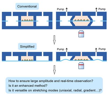
Figure 1. Schematic presentation of a simplified versus conventional microfluidic chip for cell stretching. While the conventional microfluidic chip consists of three layers (called the “three-layer chip”), the simplified one consists of only two layers (called the “two-layer chip”).
The possible bottleneck comes from varying the focus plane in z-direction during stretching driven by negative pressure, which must be significant under large stretching ratios. Ding’s group overcame such difficulty to a large extent, in the case of appropriate device parameters which were determined based upon finite element analysis and orthogonal experimental design. The novel chip was fabricated and confirmed to work in frequency up to 2 Hz and stretching ratio up to 20%.
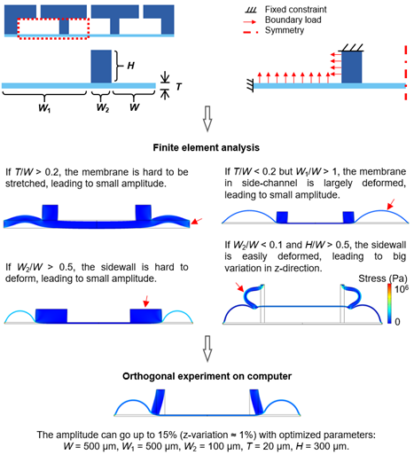
Figure 2. Optimization of chip structure by finite element analysis and orthogonal experimental design. In order to facilitate the calculation of the physical model, the chip structure was refined to obtain simple geometric structures and boundary conditions, and then the deformation of the chip under the action of air pressure was studied by finite element analysis. Finally, the optimal structure was obtained on computer through orthogonal experimental design. The color scale of the simulation outputs represents the distribution of stress. W1: width of activation cavity, W2: width of sidewall, T: thickness of membrane, H: height of microchannel.
Ding’s group further explored the extendibility of the two-layer chip for multiple stretching modes (uniaxial, radial, and gradient). Besides achieving the uniaxial stretching, the chips of radial stretching and gradient stretching were further fabricated with facile photomask design and fabrication processes. By carefully comparing the cell orientation with the displacement field for each stretching mode, we found that the orientation of cells is perpendicularly correlated to the displacement field. Since the displacement represents the disturbance to the cells, it is reasonable that individual cells oriented perpendicular to the local displacement, that is, the direction with least disturbance.
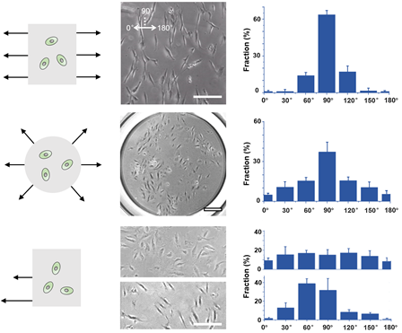
Figure 3. The three stretching modes and their cell experimenting results. The items shown from left to right are the stretching mode, cell states, and statistics of cell orientation. Scales bars: 200 μm.
The two-layer chip enables a close observation of cytoskeleton through a short working distance (~20 μm, i.e., the membrane thickness), simply owing to the removal of the third layer, which otherwise appear in the conventional three-layer chips. Both cytoskeletons changed the orientation. There are significant differences in orientation (i.e., order parameter) before and after stretching. After stretching, both cytoskeletons showed high order of orientation (Figure 4).
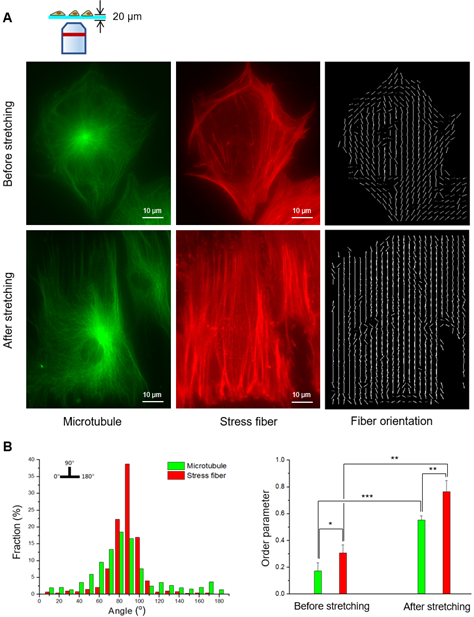
Figure 4. Fluorescence micrographs of cytoskeleton structures and their statistics. (A) Fluorescence micrographs of microtubules (left column) and stress fibers (middle column) before and after uniaxial stretching, and the fiber orientation marked by computer programs developed by the authors. (B) The left figure shows the orientation statistics of microtubule and stress fiber after uniaxial stretching. The right figure shows the order parameters of the cytoskeleton before and after uniaxial stretching. Marked in the figure are significant differences with “*” (p < 0.05), “**” (p < 0.01) and “***” (p < 0.001)
Ding’s group developed a simplified yet enhanced and versatile microfluidic platform for cell stretching. The novel two-layer chip could, albeit simplified, be a versatile and powerful tool in studies of cell mechanics and biomedical materials.
The article was published in the journal of Biofabrication with Dr. Yingning He as the first author and Professor Jiandong Ding as corresponding author. See please: Yingning He, Tianjiao Mao, Yexin Gu, Yuqian Yang, Jiandong Ding*, A simplified yet enhanced and versatile microfluidic platform for cyclic cell stretching on an elastic polymer. Biofabrication, 12 (2020) 045032.
Article link: https://iopscience.iop.org/article/10.1088/1758-5090/abb295
Get to know us better now!

Wechat:FDUMMers
Search!
Search across our website

Revenant @ 2018 by fudan | All Rights Reserved
Powered by Weicheng